EALTD
Introduction
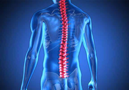
This is an endoscopic spine procedure which allows access to the postero-lateral part of the spine. A major source of back and leg pain originates from pinching of the nerves in the foramen or opening created by adjacent vertebrae. This area is accessed for surgical procedures through an incision, usually of 1/4 to 1/2 inch. Decompression of the nerve is achieved by removing the disc material, bone spurs and impinging tissues.
Endoscopic spine surgery is performed for the purpose of reducing tissue trauma, preventing iatrogenic spine problems and also for preserving spinal motion as well as stability.
The most obvious advantages of endoscopic procedures over open surgery are:
1. Smaller incisions and less surgical tissue trauma
2. Improved illumination and visibility
3. Minimal blood loss
We have all the information you need about public and private clinics and hospitals that provide spinal surgery in Iran, Islamic Republic Of with the best quality and lowest possible prices
4. Earlier return to work and daily activities
5. Easier operative option in obese patients
6. Revision surgery is easier if needed as there is less scar tissue in the access port
7. Lower rate of complications
Endoscopic strategies have been and are being performed predominantly for the treatment of the conditions such as lumbar, thoracic and cervical disc herniations with radicular pain symptoms.
However, before performing any of these procedures, a clear clinical picture of the condition obtained from complete patient history and a thorough physical as well as neurological examination is essential.
The degenerative conditions of intervertebral disc and spine are very common as evidenced by frequency of these findings on radiographs and magnetic resonance imaging (MRI). Thus, imaging studies alone can be very misleading if the pathological findings on such studies are not clearly correlated to clinical symptoms of the patient.
Endoscopic spine surgery is performed for the purpose of reducing tissue trauma, preventing iatrogenic spine problems and also for preserving spinal motion as well as stability.
When suspecting cervical as well as with lumbar spinal pain syndromes, the clinical examination should also cover the shoulder girdle and the upper extremity or the pelvis, the sacroiliac joint and the hip joints, respectively. This is important as many times, the painful conditions in these adjacent regions can mimic the symptoms caused by spinal conditions.
In equivocal situations, the use of fluoroscopy-guided, contrast-enhanced diagnostic injections is recommended to ascertain a diagnosis that is amenable to endoscopic spinal surgery.
Adequate and recent diagnostic imaging studies such as spinal MRI or computed tomography (CT) are required. In cases with changing symptoms, a repeat study prior to surgery is recommended.
Neurophysiologic studies such as electromyography or nerve conduction studies may be helpful if the diagnosis of radicular lesion is still uncertain based on patient history, clinical examination and imaging studies.
Diagnostic tests
1. Plain Radiographs – These are still considered a standard for 2 reasons. One is, they allow a quick assessment of spinal alignment, bone integrity and spine instabilities. The other is, they also allow detection of transitional vertebrae in situations where radicular pain symptoms do not match the level of an affected nerve root and thus prevent wrong-level surgery.
2. Functional myelography– injecting a dye in CSF and taking X-rays of spine is still an extremely valuable study.
3. Computed Tomography (CT) -There are some diagnostic situations where CT still is of importance, especially when MRI is not applicable (e.g.: in patients with pacemakers). CT scan allows for the reconstruction of alternative planes from the original data set, which may be of help in assessing foraminal problems which are causing symptoms. Whenever an MRI cannot be performed, post-myelography CT is an extremely valuable imaging study for evaluating disc problems.
4. MRI – This is still a gold-standard imaging study for spinal disc herniation. One of such endoscopic procedure is the selective Endoscopic Discectomy.
Procedure:
1. The patient is positioned on the operating room table in a face down position padded by special pillows. The procedure can be performed under local anesthesia.
2. After a local anesthetic is administered, a small needle is inserted into the disc space. A skin incision of about 7mm (1/4-inch) is made and a slightly larger probe is slipped over the needle into the abnormal disc.
3. Using fluoroscopic or endoscopic guidance, micro-instruments such as mini forceps, curettes, cutters, radiofrequency devices, and a laser probe are inserted.
4. The damaged parts of the disc and any bone spurs are removed. The laser is used to further remove and shrink the disc (decompression) and to tighten the annulus.
5. Exposure of the patient to x-ray is minimal. On average, the procedure takes about 30 minutes to one-hour per disc.
6. The supporting structure of the disc is not affected in this procedure. Upon completion of the surgery, the probe is removed and a small bandage is applied over the needle incision.